Three-dimensional printing has taken the world by storm. It is now possible to create any solid three-dimensional object from its digital image. Till now, we have seen a lot of its applications in automobiles, construction, technology and robotics. However, it is in medicine that 3D printing offers some exciting possibilities, be it in creating 3D models to conduct surgeries or using custom-made implants for patients.
Doctors in India are not lagging behind when it comes to trying their hand at possibilities of 3D printing technology. Here are two such attempts in Mumbai which bring hope for the future of medicine.
Facial correction
It took three years and the marvels of 3D technology for Gwalior-based Subhash Chandra Gupta to look the same as he once did. 45-year-old Gupta, diagnosed with oral cancer three years ago, had to undergo a surgical removal of left side of his jaw.
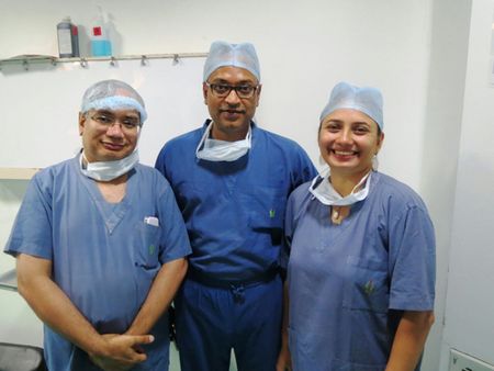
 Guruprasad Rao of Imaginarium, oncosurgeon Dr Shishir Shetty and maxillofacial prosthodontist Dr Manali Pankaj Mehta
While it helped reign in the spread of cancer, it led to major disfigurement of his face. Post the life-saving surgery, Gupta had difficulty eating and talking and was desperate for a solution. Last week, doctors from Fortis Hospital, Vashi, created a 3D-printed titanium implant of the mandible plate custom made for Gupta. Oncosurgeon Dr Shishir Shetty who conducted the surgery says conventionally patients like Gupta are fitted with a metal plate or a bone graft from his leg. Since the implants don’t fit perfectly as required, the outcomes are not satisfactory. “Using 3D printing, we could customise and make precise a imprint for the patient, which helped him look almost the same as he did before.”
“Oral cancer patients usually come to us asking to fix their jaws. But since there is no bone or support, we cannot fix their dental implants,” says Dr Manali Pankaj Mehta, consulting maxillofacial prosthodontist at Fortis Hospital. “Usually it takes two surgeries, first the oral surgery to remove the cancer affected area and second to implant the prosthesis. This takes more time and money,” says Mehta.
To fix Gupta’s problem, Mehta and Shetty consulted Imaginarium, an Andheri-based 3D printing firm. They first created a model in nylon and decided how the implant would be fixed. “We created a titanium mandible implant for the patient since it is light in weight and is not rejected by the patient,” says Shetty. Due to the precision of the technology, the doctors could fix the dentures and mandible in one sitting instead of two separate surgeries. The procedure which Mehta described as 'fairly simple and precise' took only an hour and took care of the disfigurement. It also improved Gupta's speech and eating abilities.
“This technology is brilliant and can be applied to fix any kind of defect in the bone—be it in facial surgery, neurosurgery or even oral surgery—as it takes the exact shape of the required part,” says Shetty who is now hopeful of conducting more such surgeries in the future.
Precision in surgery
When paediatric cardiac surgeon Dr Vijay Agarwal conducts a surgery, he wants to know exactly what to expect. Sometimes, in spite of the best imaging technologies like ECHO cardiogram and CT scans, he discovers that the procedure that the team prepared for cannot be conducted or needs to be altered.
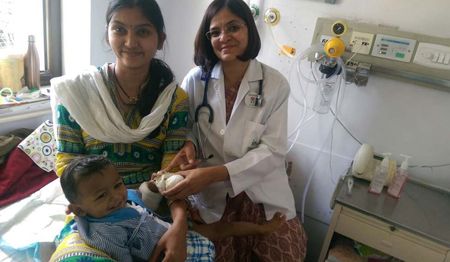
 Dr Swati Garekar with the 10-month-old baby who underwent cardiac surgery
“These kind of surprises can prove to be life-threatening for the patient,” he says. This is why when his colleague paediatric cardiologist Dr Swati Garekar learnt how 3D heart models can be used in cardiac surgeries at the Asia Pacific heart Conference in Delhi, she was excited. “We knew that studying a 3D heart model of the patient will help us plan our procedures precisely, hence, improving outcomes.”
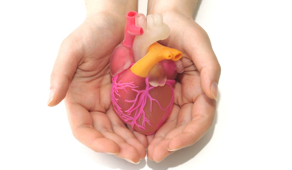
With the help of Dr Alpa Bharati who works with the radiology department at state-run Sion hospital, they created life-size heart models in five cases of patients with cardiac anomaly of double outlet right ventricle. The doctors used MRI and CT scan images to recreate the 3D sandstone heart models of infants so that cardiac surgeons could get an accurate picture and feel of what they will find during the surgery.
Agarwal who conducted the surgery on a ten-month-old baby last month says studying 3D model makes the procedure less risky and reduces the chances of patient mortality. While it takes coordination of radiology, cardiology and surgical teams and plenty of time to create the models, the results are worth it. “This is the first time a 3D heart model has been used in India. Even in the US, this kind of a model was used only in July last year. We are hoping doctors here will accept 3D technology and tap its immense benefits,” says Agarwal.


